NCERT Solution Human Reproduction Class 12 Chapter 3 Explanation Question and Answer
Human Reproduction Class 12 taken from NCERT Book Biology Chapter 3 there is all the topics are covered According to CBSE Syllabus Read and clear your all the doubts.
Human Reproduction Class 12 Introduction
Human Reproduction is a complex and intricate biological process responsible for the continuation of the human species. In Class 12 biology, the study of human reproduction delves into the anatomical, physiological, and hormonal aspects of the male and female reproductive systems. Topics include gametogenesis, fertilization, embryonic development, and the hormonal regulation of reproductive functions. Understanding human reproduction is essential not only for comprehending the biological basis of life but also for addressing reproductive health, fertility, and related societal aspects.
Human Reproduction Class 12 Characters of the Chapter
- Male Reproductive System:
- Testes: Primary male reproductive organs producing sperm and testosterone.
- Epididymis: Site of sperm maturation and storage.
- Vas Deferens: Tube connecting the epididymis to the urethra for sperm transport.
- Seminal Vesicles, Prostate Gland, Bulbourethral Glands: Accessory glands producing seminal fluid.
- Female Reproductive System:
- Ovaries: Primary female reproductive organs producing eggs (ova) and hormones (estrogen and progesterone).
- Fallopian Tubes: Tubes where fertilization typically occurs.
- Uterus: Organ where the embryo implants and develops during pregnancy.
- Cervix and Vagina: Structures involved in childbirth and part of the birth canal.
- Gametogenesis:
- Spermatogenesis: Process of sperm formation in males.
- Oogenesis: Process of egg formation in females.
- Menstrual Cycle:
- Follicular Phase, Ovulation, Luteal Phase: Phases of the menstrual cycle regulated by hormones.
- Fertilization:
- Union of Sperm and Egg: Formation of a zygote, typically occurring in the fallopian tubes.
- Embryonic Development:
- Cleavage, Morula, Blastula: Early stages of embryonic development.
- Gastrulation, Neurulation: Formation of germ layers and neural tissue.
- Pregnancy and Birth:
- Implantation: Attachment of the embryo to the uterine wall.
- Placenta: Organ facilitating nutrient and gas exchange between mother and fetus.
- Labor and Delivery: Process of childbirth.
- Reproductive Health:
- Contraception and Family Planning: Methods to control fertility.
- Sexually Transmitted Infections (STIs): Health risks associated with unprotected sexual activity.
- Infertility: Challenges and treatments related to difficulty in conceiving.
Human Reproduction Class 12 Definition
Human Reproduction involves the study of the biological processes essential for the continuation of the human species. It encompasses the anatomical, physiological, and hormonal aspects of the male and female reproductive systems, gametogenesis (formation of gametes), fertilization, embryonic development, and the intricate hormonal regulation of reproductive functions. This chapter provides insights into the mechanisms underlying human reproduction, contributing to a comprehensive understanding of the complexities associated with the creation and development of new life.
Human Reproduction Class 12 Explanaion
As you are aware, humans are sexually reproducing and viviparous. The reproductive events in humans include formation of gametes (gametogenesis), i.e., sperms in males and ovum in females, transfer of sperms into the female genital tract (insemination) and fusion of male and female gametes (fertilisation) leading to the formation of zygote. This is followed by the formation and development of blastocyst and its attachment to the uterine wall
Meaning of this word (fertilisation): “Fertilization is the process in sexual reproduction where a sperm cell fuses with an egg cell, resulting in the formation of a zygote.”
(implantation), embryonic development (gestation), and delivery of the baby (parturition). You have learnt that these reproductive events occur after puberty. There are remarkable differences between the reproductive events in the male and in the female, for example, sperm formation continues even in old men, but the formation of ovum ceases in women around the age of fifty years. Let us examine the male and female reproductive systems in humans.
3.1 THE MALE REPRODUCTIVE SYSTEM
The male reproduction system is located in the pelvis region (Figure 3.1a) it includes a pair of testes alongwith accessory ducts, glands and the external genitalia.
The testes are situated outside the abdominal cavity within a pouch called the scrotum. The scrotum helps in maintaining the low temperature of the testes (2–2.5o C lower than the normal internal body temperature) necessary for spermatogenesis. In adults, each testis is oval in shape, with a length of about 4 to 5 cm and a width of about 2 to 3 cm. The testis is covered by a dense covering

Meaning of this word spermatogenesis : “Spermatogenesis is the process of sperm cell formation in the male reproductive system through meiotic divisions and maturation.”
.Each testis has about 250 compartments called testicular lobules (Figure 3.1b). Each lobule contains one to three highly coiled seminiferous tubules in which sperms are produced. Each seminiferous tubule is lined on its inside by two types of cells called male germ cells
(spermatogonia) and Sertoli cells (Figure 3.2 ). The male germ cells undergo meiotic divisions finally leading to sperm formation, while Sertoli cells provide nutrition to the germ cells. The regions outside the seminiferous tubules called interstitial spaces, contain small blood vessels and interstitial cells.
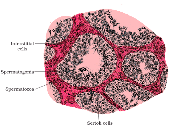
or Leydig cells (Figure 3.2). Leydig cells synthesize and secrete testicular hormones called androgens. Other immunologically competent cells are also present. The male sex accessory ducts include rete testis, vasa efferentia, epididymis, and vas deferens (Figure 3.1b). The seminiferous tubules of the testis open into the vasa efferentia through rete testis.
The vasa efferentia leave the testis and open into the epididymis located along the posterior surface of each testis. The epididymis leads to the vas deferens that ascends to the abdomen and loops over the urinary bladder. It receives a duct from the seminal vesicle and opens into the urethra as the ejaculatory duct (Figure 3.1a).
These ducts store and transport the sperms from the testis to the outside through the urethra. The urethra originates from the urinary bladder and extends through the penis to its external opening called urethral meatus.
The penis is the male external genitalia (Figure 3.1a, b). It is made up of special tissue that helps in the erection of the penis to facilitate insemination. The enlarged end of the penis called the glans penis is covered by a loose fold of skin called the foreskin. The male accessory glands (Figure 3.1a, b) include paired seminal vesicles, a prostate, and paired bulbourethral glands. Secretions of these glands constitute the seminal plasma, which is rich in fructose, calcium, and certain enzymes. The secretions of the bulbourethral glands also help in the lubrication of the penis.
Meaning of this word bulbourethral glands:”Bulbourethral glands are small structures in the male reproductive system that produce a clear lubricating fluid, contributing to semen.”
3.2 THE FEMALE REPRODUCTIVE SYSTEM
The female reproductive system consists of a pair of ovaries along with a pair of oviducts, uterus, cervix, vagina, and the external genitalia located in the pelvic region (Figure 3.3a). These parts of the system along with a pair of mammary glands are integrated structurally and functionally to support the processes of ovulation,
fertilization, pregnancy, birth, and child care. Ovaries are the primary female sex organs that produce the female gamete (ovum) and several steroid hormones (ovarian hormones). The ovaries are located one on each side of the lower abdomen (Figure 3.3b). Each ovary is about 2 to 4 cm in length and is connected to the pelvic wall and uterus by ligaments. Each ovary is covered by a thin epithelium that encloses the ovarian stroma. The stroma is divided into two zones – a peripheral cortex and an inner medulla.
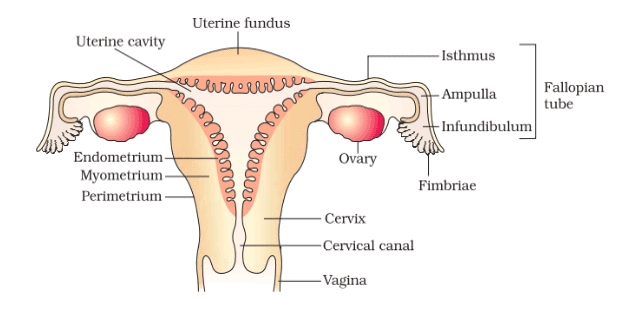
The oviducts (fallopian tubes), uterus, and vagina constitute the female accessory ducts. Each fallopian tube is about 10-12 cm long and extends from the periphery of each ovary to the uterus (Figure 3.3b), the part closer to the ovary is the funnel-shaped infundibulum. The edges of the infundibulum possess finger-like projections called fimbriae, which help in the collection of the ovum after ovulation. The infundibulum leads to a wider,
Part of the oviduct called ampulla. The last part of the oviduct, isthmus has a narrow lumen and it joins the uterus. The uterus is single and it is also called womb. The shape of the uterus is like an inverted pear. It is supported by ligaments attached to the pelvic wall.
The uterus opens into the vagina through a narrow cervix. The cavity of the cervix is called cervical canal (Figure 3.3b) which along with the vagina forms the birth canal. The wall of the uterus has three layers of tissue. The external thin membranous perimetrium, middle thick layer of smooth muscle, myometrium
and inner glandular layer called endometrium that lines the uterine cavity. The endometrium undergoes cyclical changes during the menstrual cycle while the myometrium exhibits strong contractions during the delivery of the baby. The female external genitalia include mons pubis, labia majora, labia minora,
hymen, and clitoris (Figure 3.3a). Mons pubis is a cushion of fatty tissue covered by skin and pubic hair. The labia majora are fleshy folds of tissue, which extend down from the mons pubis and surround the vaginal opening. The labia minora are paired folds of tissue under the labia majora. The opening of the vagina is often covered partially by a membrane called hymen. The clitoris is a tiny finger-like structure that lies at the upper junction of the two labia minora above the urethral opening.
The hymen is often torn during the first coitus (intercourse). However, it can also be broken by a sudden fall or jolt, insertion of a vaginal tampon, active participation in some sports like horseback riding, cycling, etc. In some women, the hymen persists even after coitus. In fact, the presence or absence of the hymen is not a reliable indicator of virginity or sexual experience.
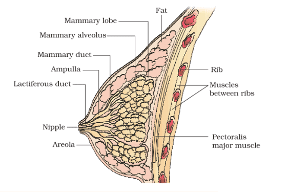
A functional mammary gland is characteristic of all female mammals. The mammary glands are paired structures (breasts) that contain glandular tissue and a variable amount of fat. The glandular tissue of each breast is divided into 15-20 mammary lobes containing clusters of cells called alveoli (Figure 3.4). The cells of alveoli secrete milk,
which is stored in the cavities (lumens) of alveoli. The alveoli open into mammary tubules. The tubules of each lobe join to form a mammary duct. Several mammary ducts join to form a wider mammary ampulla which is connected to the lactiferous duct through which milk is sucked out.
3.3 GAMETOGENESIS
The primary sex organs – the testis in males and the ovaries in females – produce gametes, i.e., sperms and ovum, respectively, by the process called gametogenesis. In the testis, the immature male germ cells (spermatogonia) produce sperms by spermatogenesis that begins at puberty. The spermatogonia (sing. spermatogonium) present on the inside wall of seminiferous tubules multiply by mitotic division and increase in numbers. Each
spermatogonium is diploid and contains 46 chromosomes. Some of the spermatogonia called primary spermatocytes periodically undergo meiosis. A primary spermatocyte completes the first meiotic division (reduction division) leading to the formation of two equal, haploid cells called secondary spermatocytes, which have only 23 chromosomes each. The secondary spermatocytes undergo the second meiotic division to produce four equal
haploid spermatids (Figure 3.5). What would be the number of chromosomes in the spermatids? The spermatids are transformed into spermatozoa (sperms) by the process called spermiogenesis. After spermiogenesis, sperm heads become embedded in the Sertoli cells and are finally released from the seminiferous tubules by the process called spermiation.
Spermatogenesis starts at the age of puberty due to a significant increase in the secretion of gonadotropin-releasing hormone (GnRH). This, if you recall, is a hypothalamic hormone. The increased levels of GnRH then act at the anterior pituitary gland and stimulate the secretion of two gonadotropins – luteinizing hormone (LH) and follicle-stimulating hormone (FSH). LH acts at the Leydig cells and stimulates the synthesis and secretion of androgens. Androgens, in turn, stimulate the process of spermatogenesis. FSH acts on the Sertoli cells and stimulates.
Human Reproduction Class 12 Important Notes
- Gametogenesis:
- Gametogenesis is the process of gamete formation, with spermatogenesis occurring in the testes of males and oogenesis taking place in the ovaries of females.
- Spermatogenesis begins at puberty and continues throughout life, while oogenesis begins before birth but is only completed after puberty.
- Hormonal Regulation:
- Gonadotropin-releasing hormone (GnRH) from the hypothalamus stimulates the release of luteinizing hormone (LH) and follicle-stimulating hormone (FSH) from the pituitary gland.
- LH stimulates androgen production in males (by Leydig cells) and ovulation in females, while FSH promotes spermatogenesis in males and follicular development in females.
- Menstrual Cycle:
- The menstrual cycle is a recurring physiological process in females, involving the preparation of the uterus for a potential pregnancy.
- Phases include menstruation, the follicular phase (pre-ovulatory), ovulation, and the luteal phase (post-ovulatory).
Human Reproduction Class 12 summary
Humans are sexually reproducing and viviparous. The male reproductive system is composed of a pair of testes, the male sex accessory ducts, and the accessory glands and external genitalia. Each testis has about 250 compartments called testicular lobules, and each lobule contains one to three highly coiled seminiferous tubules.
Each seminiferous tubule is lined inside by spermatogonia and Sertoli cells. The spermatogonia undergo meiotic divisions leading to sperm formation, while Sertoli cells provide nutrition to the dividing germ cells. The Leydig cells outside the seminiferous tubules synthesize and secrete testicular hormones called androgens
The male external genitalia is called the penis. The female reproductive system consists of a pair of ovaries, a pair of oviducts, a uterus, a vagina, external genitalia, and a pair of mammary glands. The ovaries produce the female gamete (ovum) and some steroid hormones (ovarian hormones). Ovarian follicles in different stages of development are embedded in the stroma. The oviducts, uterus
and vagina are female accessory ducts. The uterus has three layers namely perimetrium, myometrium, and endometrium. The female external genitalia includes mons pubis, labia majora, labia minora, hymen, and clitoris. The mammary glands are one of the female secondary sexual characteristics.
Spermatogenesis results in the formation of sperms that are transported by the male sex accessory ducts. A normal human sperm is composed of a head, neck, a middle piece, and tail. The process of the formation of mature female gametes is called oogenesis. The reproductive cycle of female primates is called the menstrual cycle.
The menstrual cycle starts only after attaining sexual maturation (puberty). During ovulation, only one ovum is released per menstrual cycle. The cyclical changes in the ovary and the uterus during the menstrual cycle are induced by changes in the levels of pituitary and ovarian hormones. After coitus, sperms are transported to the junction of the isthmus and ampulla, where the sperm fertilizes the ovum leading to the formation of a diploid zygote.
The presence of X or Y chromosome in the sperm determines the sex of the embryo. The zygote undergoes repeated mitotic division to form a blastocyst, which is implanted in the uterus resulting in pregnancy. After nine months of pregnancy, the fully developed fetus is ready for delivery. The process of childbirth is called parturition which is induced by a complex neuroendocrine mechanism involving cortisol.
Human Reproduction Class 12 Question and Answer
Question:1 Define spermiogenesis and spermiation
Answer
Spermiogenesis: Spermiogenesis is the process of maturation and differentiation of spermatids into functional spermatozoa (sperms) in the seminiferous tubules of the testes. During this process, the round-shaped spermatids undergo structural and functional changes, including the development of a tail, which transforms them into mature sperm cells capable of fertilization.
Spermiation: Spermiation is the final step in spermatogenesis, during which mature spermatozoa are released from the Sertoli cells lining the seminiferous tubules into the lumen of the tubules. This allows the mature sperm cells to enter the epididymis, where they undergo further maturation and acquire the ability to swim and fertilize an egg during sexual reproduction.
Question:2 What are the major components of seminal plasma?
Answer:
- Fructose: Provides energy for sperm metabolism.
- Prostaglandins: Regulate the female reproductive tract to facilitate sperm movement.
- Enzymes: Aid in breaking down substances in the female reproductive tract, facilitating sperm penetration.
- Buffers: Help maintain the optimal pH for sperm function.
- Zinc: Important for sperm function.
- Calcium: Essential for sperm motility.
- Immunoglobulins: Provide some protection against infections.
- Citric Acid: Contributes to the acidity of the semen.
Question:3 What is spermatogenesis? Briefly describe the process of spermatogenesis
Answer
Process of Spermatogenesis:
- Proliferation (Mitosis): Spermatogonia, which are undifferentiated germ cells, undergo mitotic divisions to increase their numbers. Some of these cells remain as spermatogonia to maintain the germ cell pool, while others progress to the next stages.
- Meiosis I: Primary spermatocytes, resulting from the mitotic divisions, undergo the first meiotic division (meiosis I) to produce two secondary spermatocytes. This reduces the chromosome number from diploid (2n) to haploid (n).
- Meiosis II: Each secondary spermatocyte then undergoes the second meiotic division (meiosis II), resulting in the formation of four haploid spermatids. These spermatids contain half the chromosome number of the original spermatogonium.
- Spermiogenesis: Spermatids undergo a process called spermiogenesis, during which they undergo structural and morphological changes. The round spermatids transform into mature spermatozoa with a head, midpiece, and tail.
- Spermiation: Mature spermatozoa are released from the Sertoli cells into the lumen of the seminiferous tubules through a process called spermiation.
Human Reproduction Class 12 MCQ Question and Answer
Question:1. Which hormone stimulates the Leydig cells to produce androgens in males?
- A) Follicle-stimulating hormone (FSH)
- B) Luteinizing hormone (LH)
- C) Gonadotropin-releasing hormone (GnRH)
- D) Testosterone
- Answer: B) Luteinizing hormone (LH)
Question:2. Where does fertilization typically occur in the female reproductive system?
- A) Uterus
- B) Oviduct (Fallopian tube)
- C) Vagina
- D) Cervix
- Answer: B) Oviduct (Fallopian tube)
Question:3. What is the primary function of the seminal vesicles in male reproductive anatomy?
- A) Sperm production
- B) Hormone secretion
- C) Secretion of alkaline fluid
- D) Lubrication of the penis
- Answer: C) Secretion of alkaline fluid
Question:4. What is the function of the corpus luteum during the menstrual cycle?
- A) Production of estrogen
- B) Secretion of progesterone
- C) Ovulation induction
- D) Formation of ova
- Answer: B) Secretion of progesterone
Question:5. Which structure connects the testis to the urethra and transports sperm?
- A) Vas deferens
- B) Epididymis
- C) Seminal vesicle
- D) Prostate gland
- Answer: A) Vas deferens
Question:6. What is the role of Sertoli cells in the male reproductive system?
- A) Sperm production
- B) Androgen synthesis
- C) Nutrition for germ cells
- D) Lubrication of the penis
- Answer: C) Nutrition for germ cells
Question:7. Which part of the female reproductive system is responsible for the development of the blastocyst before implantation?
- A) Uterus
- B) Ovary
- C) Cervix
- D) Vagina
- Answer: A) Uterus
Question:8. What is the purpose of the cervical mucus in the female reproductive system?
- A) Nourishment for ova
- B) Lubrication
- C) Protection against infections
- D) Facilitation of sperm entry
- Answer: D) Facilitation of sperm entry
Question:9. What triggers the release of GnRH (Gonadotropin-releasing hormone) during the menstrual cycle?
- A) High estrogen levels
- B) Low progesterone levels
- C) Low estrogen levels
- D) High progesterone levels
- Answer: C) Low estrogen levels
Question: 10. Which structure is responsible for the production of ova in the female reproductive system?
- A) Uterus
- B) Ovary
- C) Fallopian tube
- D) Vagina
- Answer: B) Ovary
Conclusion
Human Reproduction explores the intricate processes of gametogenesis, fertilization, and embryonic development, offering a profound understanding of the complexities underlying human reproduction.